
UReCA: The NCHC Journal of Undergraduate Research and Creative Activity 2020 Edition
Natural Oils Protect Human Skin Cells against Harmful UVB Radiation
ABSTRACT
Ultraviolet (UV) radiation from the sun is a recognized cause of skin cancer, which affects millions of people each year around the world. The chemopreventative efficacy of canola and olive oils was tested against UVB-irradiation-induced death and DNA damage in human immortalized keratinocytes (HaCaT) from adult human skin. The study revealed that both oils are safe for human application, as they were non-toxic to the HaCaT. The UVB protective efficacy was monitored by first coating the bottom of the petri dishes (containing growing HaCaT) with canola and olive oils and then irradiating them from below with UV light. This indicates that the oils could effectively block the penetration of UVB to the HaCaT. Additionally, the HaCaT were pretreated with canola and olive oil before irradiation in another test, and significant protection was found, suggesting that the oils are not only photoprotective but also have biological activity. To understand the mechanism behind the protective efficacy of the oils, the cyclobutane-pyrimidine-dimer (CPD) formation, or DNA damage, was examined. Analysis showed that the UVB-treated cells had high CPD formation, while cells pretreated with oils had significantly low CPD formation as revealed by a dot-blot assay. Together, these findings provide support for the utility of canola and olive oils as potential sunscreen agents or additives.
KEYWORDS
Ultraviolet; Skin Cancer; Natural; Safe; Oil; DNA; HaCaT; Sunscreen; Nanoparticles
Introduction
Every year in the United States, there are more new cases of skin cancers, such as melanoma, than combined incidences of cancers of the breast, prostate, lung and colon. According to the Skin Cancer Foundation, almost 86 percent of melanomas are caused in part by exposure to ultraviolet (UV) radiation from the sun.1
Many people use sunscreen lotions to protect themselves from harmful UV radiation. However, these protective agents have failed to curb the incidence of skin cancer in the United States.2 Additionally, about nineteen percent of the U.S. population has reported allergic reactions to active ingredients such as titanium dioxide and zinc oxide in major sunscreens. Previous research has shown these ingredients to be harmful to the body in concentrations greater than approximately 22 percent of the entire sunscreen when applied to the skin in a 2 mg/cm2 layer.3 Several popular sunscreens have zinc oxide concentrations at these dangerous levels, such as UV Natural Sport Sunscreen SPF 30+ with a zinc oxide concentration of 24.8%, despite being advertised as a “natural” sunscreen.4
Alternative forms of natural sun protection exist in civilizations around the world. For example, applying coconut oil to the skin is a common practice in the tropical Philippines, located within the sunny Tropic of Cancer. Coconut oil blocks approximately 20% of the sun’s rays, contributes to healthy vitamin D production, and can also be easily found in household kitchens.5 Having overlooked this common household oil, the experimenter explored what other common oils could be tested for natural sunscreen protection. Olive oil is commonly used to cook foods in Italy and Spain and was also bathed in by Egyptian Pharaoh Cleopatra to revitalize her skin.6 Both oils are staple moisturizers amongst beauty circles worldwide. The experimenter planned on testing these oils to determine the validity of these anecdotal claims. An additional oil planned for testing was canola oil, which is not commonly associated with UV protection. On the other hand, its dark color and high viscosity piqued curiosity make one consider the possibility of using it as a sunscreen agent. Current research is lacking exploration into these common kitchen oils, though seemingly exotic oils such as jojoba oil, carrot seed oil, soybean oil, etc. have already been proposed.14 Successful experimentation with easily accessible oils could potentially have a significant impact on what sunscreens are sold commercially in the future.
If a natural oil could be used in combination with another active ingredient such as silver nanoparticles7, it could be used to create a potentially worthwhile sunscreen compound. Out of pure coconut oil, olive oil, and canola oil, which substance would protect the greatest number of HaCaT from death and result in its protected cells having the highest percent viability?
METHODS AND PROCEDURES
Culturing HaCaT
HaCaT were cultured in ventilated flasks inside a biosafety cabinet at the USA Mitchell Cancer Institute in Mobile, Alabama. Cells were maintained in a humidified atmosphere of 5% CO2 at 37°C as monolayer culture in DMEM medium supplemented with 10% Fetal Bovine Serum (FBS), penicillin (100 units/mL), and streptomycin (100 µL/mL). Cells were cultured and maintained in accordance with the Centers for Disease Control Biosafety Level 2.8
Testing in vitro SPF of the oils
1 μL of each oil was kept on the UV Nanodrop ND-1000 Spectrophotometer and light in the UVB spectrum (290 to 320 nm) was shone through each oil to give each sample an absorbance reading of 0-4, with 4 being the highest amount of UV light absorbed and 0 being the lowest. Using the Mansur Equation9 and UVB constants created by Sayre et. al (1979), the experimenter converted the absorbance values for each wavelength into an in vitro Sun Protection Factor. Since canola oil had the highest SPF, it was predicted that it would be the most effective carrier oil in the hypothesis. Due to coconut oil’s low SPF, it was not tested in the following experiments.

Toxicity evaluation
Each oil was tested for toxicity in a 96-well, treated microplate by seeding an equal amount of HaCaT in each well and mixing in DMEM and canola/olive oils in different concentrations. This toxicity evaluation determined if either of the oils were toxic to the HaCaT, and if so, in what concentrations were they the most toxic. Concentrations tested include 0, 5, 10, 25, 50, and 100 mg/mL ratio of coconut/olive oil to DMEM. A Perkin Elmer UV spectrophotometer was used to quickly measure each well’s percent viability. The percent viability of each well was monitored after 6, 12, 24, and 48 hours to determine if the oils decreased cell viability over extended periods of time. Testing toxicity before experimentation was essential to make certain that cells died from UV exposure during trials and not from exposure to oils. Additionally, if oils were highly toxic to HaCaT, experimentation would have to be halted as neither coconut nor olive oil would be suitable bases for a potential sunscreen.

Two procedures required the use of a UV Transilluminator: the coating test and the pretreatment test.
Coating tests
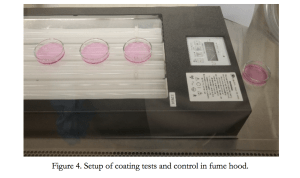
Two procedures required the use of a UV Transilluminator: the coating test and the pretreatment test. The coating test was an indirect method of protecting the cells. In essence, it consisted of evaluating the percent viability of the cells seeded in petri dishes before and after UVB irradiation. Coconut oil and canola oil covered the bottom of two petri dishes in a 2 mg/cm2 ratio as this is the amount commercial companies use in clinical sunscreen trials with live patients per FDA regulation. Additionally, there were two “control” petri dishes, one with no treatment at all and one with only UVB treatment, creating a total of four petri dishes. These petri dishes were then placed on the UV Transilluminator at least 3 cm apart from each other, and 40 mJ of UVB light was emitted from below the dishes. 40 mJ of UVB is the typical amount of radiation emitted by the sun on an average summer day, which justifies this setting in the irradiation experiments. After the dishes were exposed to UVB, they were incubated for 24 hours to allow for the damage to become visible microscopically. Finally, the percent viability of each dish was calculated using a Countess automated cell counter. This involved dyeing a 1 µL sample of the media of each petri dish with Trypan blue dye to stain the human keratinocytes to simplify detection. These dyed samples were then transferred to cell counting chamber slides and inserted into the Countess to measure viability. This data was recorded and graphed to compare the percent viabilities of each petri dish before and after experimentation.
Pretreatment testing
The pretreatment test was a direct method of protecting the cells as it involved mixing the oils directly into the DMEM formulation from which the cells receive nutrients. Based on the toxicity evaluation, it was determined that 25 mg/mL of oil would be the largest amount possible before the oils began displaying a negative effect on cell viability. Because the natural oils and the water-based DMEM cannot mix, acetone was used as a solvent to allow emulsion to occur. They were mixed in a 1:1 ratio in a 20 mL centrifuge tube and later used as part of two stocks for distribution to the petri dishes. Since the density of both oils was 920 mg/mL, 460 mg/mL of acetone plus 460 mg/mL of canola oil was in one centrifuge tube, and 460 mg/mL of acetone plus 460 mg/mL of olive oil was in another. After this step was completed, the two stocks could be created.

This procedure was done with both canola and olive oil to create the two stocks necessary for the pretreatment tests. 3000 µL (3 mL) was used as the ideal amount of DMEM to supplement the cells in both the pretreatment tests and the coating tests. A motorized pipette was used to move 3 mL of the canola oil stock into one of four quartz petri dishes. This was repeated with the olive oil stock. Two remaining petri dishes did not receive a special stock and were simply supplemented with 3 mL of DMEM. All four petri dishes were stored in an incubator for 24 hours at 37°C before experimentation.
The remainder of the pretreatment testing was similar to the coating tests. Out of the two remaining petri dishes that were only DMEM-supplemented, one was a control dish that did not receive any treatment at all, and the other was a control dish that only received UVB treatment (no oil protection). 40 mJ of UVB light was emitted through the bottom of each petri dish, each dish was incubated for 24 hours, and then the cells were counted afterwards.
Dot-blot assay
The final experiment in the methodology of this research was the dot-blot assay, which utilized cells from the pretreatment tests. The dot-blot assay displays the intensity of DNA damage based on the formation of cyclobutane pyrimidine dimers (CPDs). Cyclobutane pyrimidine dimers are indicators of DNA damage and can be used to determine which natural oil was the most effective at preventing damage in the nucleus of the human keratinocytes. In the assay, the darker the blot, the greater the DNA damage.
500 nanograms of HaCaT genomic DNA (treated or untreated) was isolated using DNAzol and transported to a positively charged nitrocellulose membrane. The membrane was then baked for thirty minutes at 80°C to immobilize the DNA and blocked by 5% nonfat dry milk in Tris Buffered Saline with Tween 20 (TBST). The milk contains proteins which bind to the membrane to prevent the target antibody (anti-CPD antibody, in this case) from binding to any other location than the target antigen (the CPD formed). The milk prevents this non-specific binding by attaching its own phosphoproteins to open protein sites on the membrane, thus preventing the anti-CPD antibodies from binding there unintentionally. However, the binding of the anti-CPD antibody with the CPD itself is undetectable without a fluorescent secondary antibody tagging itself onto the primary antibody. After washing with TBST to remove stray proteins, the membrane was incubated with horseradish peroxidase (HRP) conjugated secondary antibody. When this fluorescent secondary antibody successfully bound to the anti-CPD primary antibody, a chemiluminescence (ECL) plus detection kit was used to detect dot-blot formation. This image was later digitized for examination using a LAS-3000 image analyzer from Fuji Photo Film Company.
RESULTS

Figure 5 displays the SPF readings given by the Nanodrop spectrophotometer in the preliminary testing. The Nanodrop gave ten absorbance readings (abs), one for each of the UVB wavelengths tested. These readings, ranging from zero to four, are shown in the second set of columns in the center of the chart. The abs for each oil was multiplied by its corresponding EE x I constant in the first column set to deliver the values shown in the third column set. When all seven of the values for each oil in the third column set were added together and multiplied by a correction factor (CF) of ten, the final in vitro SPF value for each oil was successfully calculated. The erythemal effect (EE) and solar intensity spectrum (I) were constants for each wavelength in the UVB spectrum, as shown previously in Figure 1. The Mansur equation calculation for canola oil is shown below:

The final in vitro SPF calculation for canola oil was determined to be approximately 16.51. This methodology was performed to calculate the in vitro SPF for olive oil and coconut oil.

Due to the critically low in vitro SPF of coconut oil and, it was not determined to be a viable applicant for further testing.

Figure 7 displays the results of the toxicity evaluation over a period of forty-eight hours, with incremental measurements of viability taking place after six, twelve, and twenty-four hours. As no HaCaT perished after 48 hours with no oil influence, there were no significant external factors affecting viability in this experiment. In the presence of either oil in any concentration, cell viability decreased over time, with higher oil concentrations experiencing greater reductions than lower concentrations. As sunscreens are generally not applied for over 12 hours at a time, this time frame was given the most credence during analysis. In this graph, cell viability increases with oil concentration up to 25 mg/mL, when viability begins a negative trend. Because this was found to be the most beneficial oil concentration for both canola and olive oils, this was the concentration used in the pretreatment experiments.

In the coating tests, the petri dishes that did not undergo any UVB treatment nor had any oil at the bottom of the dish remained at 100% viability. The petri dish that underwent a 40 millijoule dose of UVB radiation without any oil decreased to 60% viability. The petri dish that underwent UVB treatment with 2 mg/cm2 of canola oil coated on the bottom of the dish increased by 21% in viability when compared to the UVB treatment alone to obtain 81% viability. The petri dish that underwent UVB treatment with 2mg/cm2 of olive oil coated on the bottom of the dish increased by 16% in viability when compared to the UVB treatment alone to obtain 76%. Though there is still a notable increase in viability with the olive oil, it is not as large an increase as with the canola oil, as shown in Figure 8 above.

In the pretreatment tests, the petri dishes that did not undergo any UVB treatment nor had any oil pretreated inside of the dish remained at 100% viability. The petri dish that underwent a 40 millijoule dose of UVB radiation without any oil pretreatment decreased to 60% viability. The petri dish that underwent UVB treatment with 25 mg/mL of canola oil pretreated inside of the dish increased by 28% in viability when compared to the UVB treatment alone to obtain 88% viability. The petri dish that underwent UVB treatment with 25 mg/mL of olive oil pretreated inside of the dish increased by 22% in viability when compared to the UVB treatment alone to obtain 82% viability. Though there is still a notable increase in viability with the olive oil, it was not as large an increase as with the canola oil, as shown in Figure 9 above.

In both the coating and pretreatment tests, the petri dishes protected by olive and canola oils did not pass the eye-test for a statistically significant difference in viability. Without having a clear indication that these viabilities were indeed different, the experimenter could not confidently state that canola oil was more effective than olive oil or vice versa. Thus, an unpaired t-test was performed for both experiments as shown in Figure 10 (above) to determine if the mean viabilities of each oil were statistically different. These t-tests revealed a two-tailed P value of 0.0484 for the coating tests and 0.0434 for the pretreatment tests. As both values are below the criteria of P ≤ 0.05, these viability differences are conventionally considered to be statistically significant. Therefore, the experimenter can reasonably declare that canola oil was more effective than olive oil at protecting the human keratinocytes from death in both the coating and pretreatment experiments.
The dot-blot analysis in Figure 11 (below) assisted in the determination of the intensity of DNA damage occurring to the HaCaT after pretreatment testing. The formation of cyclobutane-pyrimidine-dimers (CPDs) indicated the level of DNA damage, with a deeper shade of black dot indicating a larger amount of DNA damage. As shown in the diagram below, the petri dish that did not undergo any UVB treatment did not obtain any DNA damage. The petri dish that did not have any oil pretreated inside of the dish but did undergo UVB treatment at 40 mJ obtained a high level of CPD formation and DNA damage. The petri dish that underwent UVB treatment at 40 mJ and had olive oil pretreated inside of the petri dish at 25 mg/mL obtained a fair level of CPD formation and UVB damage. Finally, the petri dish that underwent UVB treatment at 40 mJ and had canola oil pretreated inside of the petri dish at 25 mg/mL obtained a low level of CPD formation and UVB damage.

DISCUSSION
As shown in the results to the above, canola oil was the most effective at protecting HaCaT cells from death and DNA damage due to UVB-irradiation. Due to SPF calculation and testing, this result was expected; however, the margin between canola and olive oil in cell viability was predicted to be much greater due to the SPF of canola oil being almost twice that of olive oil. This result disproves the common myth that twice the Sun Protection Factor leads to twice the protection. It also leads to the assumption that if coconut oil had been tested, it would have yielded a similar yet slightly lower percent viability in its petri dishes. There is little variation in the data found by this experiment as both coating and pretreatment tests yielded similar results. The results of this experiment were affected by how well the cells grew and responded to their environment, so the HaCaT themselves were an inherent variable.
The pretreatment method proved to be the more effective mode of protecting the human keratinocytes than the coating method. This could be attributed to the direct nature of the pretreatment experiments compared to the indirectness of the coating method. However, it could also be attributed to the oils reacting with and triggering a biological response from the keratinocytes. Canola oil is composed of essential fatty acids such as linoleic acid and stearic acid.11 Deficiency of linoleic acid has been shown to cause mild skin scaling and hair loss in rats.12 This suggests that linoleic acid may play a role in enhancing the structural integrity of the human keratinocytes in this experiment. Stearic acid has also been shown to have antioxidant properties,13 suggesting that it could have contributed in limiting CPD formation and overall DNA damage in the keratinocytes. Implementing critical fatty acids and antioxidants such as these into commercial sunscreen products could help increase their efficacy in the future.
To enhance the validity of the results of this study, the experimenter would like to perform similar experiments with commercial sunscreens with SPFs of varying intervals of 15, 30, 50, and 100. Toxicity evaluations of modern sunscreens would determine the safest concentration that sunscreens could be exposed to keratinocytes in their medium. Pretreatment and coating tests could determine if sunscreens trigger biological activity in the keratinocytes as the oils did.
CONCLUSION
This study proposed the research question that out of pure coconut oil, olive oil, and canola oil, which substance would protect the greatest number of HaCaT from death and result in its protected cells having the highest viability. As predicted in the hypothesis, canola oil was the most effective at preventing the death and DNA damage of human keratinocytes (HaCaT) when irradiated with UVB light in coating and pretreatment tests. When tested under these rigorous experiments, canola oil reported the highest percent viability of the oils with an impressive 88% viability in pretreatment tests. Though not as effective as canola, olive oil also showed significant ability in protecting the human keratinocytes.
A future goal of this undertaking is to eventually develop a safe natural sunscreen which will use silver nanoparticles7 (AgNPs), shown by previous research to protect and repair damaged HaCaT, and a natural carrier oil. Though it is not yet advisable to use these natural oils as independent sunscreens, they could serve as effective carriers of safe active ingredients such as AgNPs, having potential to increase overall sunscreen efficacy. This research will benefit those with allergies to the zinc oxide and titanium dioxide active ingredients found in almost all major sunscreens and will also benefit those who are searching for a safe alternative to sunscreens in the market today.